
OUR TECHNOLOGY
In focus.
The instruments behind every exam at Village Vision Center — chosen because they show your doctor more, and ask less of you.
A modern exam is a series of quiet, precise measurements — here's what does the looking.
INTRAOCULAR PRESSURE
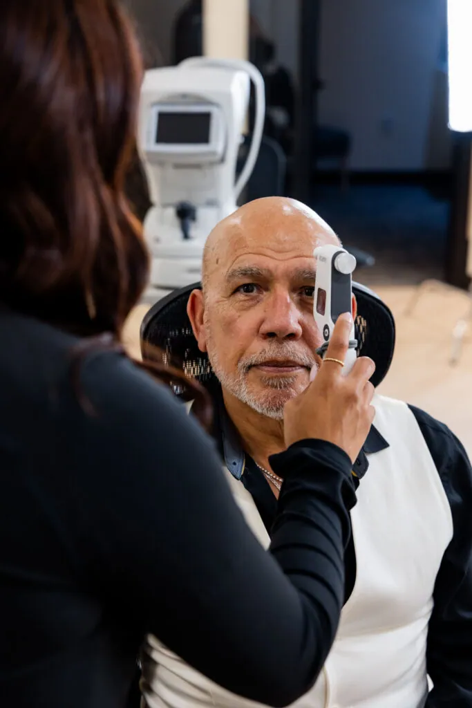
Tonometer iCare
No puff of air. No drops. A feather-light rebound probe touches the cornea for a fraction of a second to read the pressure inside your eye — an accurate screening you’ll barely notice.
-
Method Rebound
-
Contact Momentary
-
Experience Air-free · Drop-free
BASELINE PRESCRIPTION
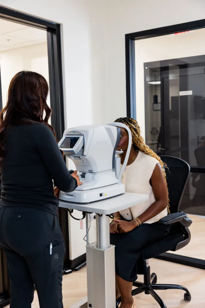
Auto-Refractor
Autorefractor and keratometer in one instrument. It measures how light bends inside your eye and maps the curvature of your cornea — giving your doctor a precise starting point before you read a single line on the chart.
-
Reads Refractive error
-
Maps Corneal curvature
-
Result Prescription baseline
Everything you can't feel, measured to the micron.
WIDE-FIELD RETINAL IMAGING
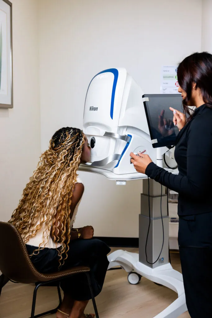
Optomap California Retinal Camera
A single capture opens a far wider view of the back of your eye than a standard retinal camera. For many patients it’s a comfortable alternative to dilation — and a richer picture for catching what matters early.
-
View Ultra-wide field
-
Comfort Dilation alternative
-
Capture Single image
CROSS-SECTIONAL IMAGING
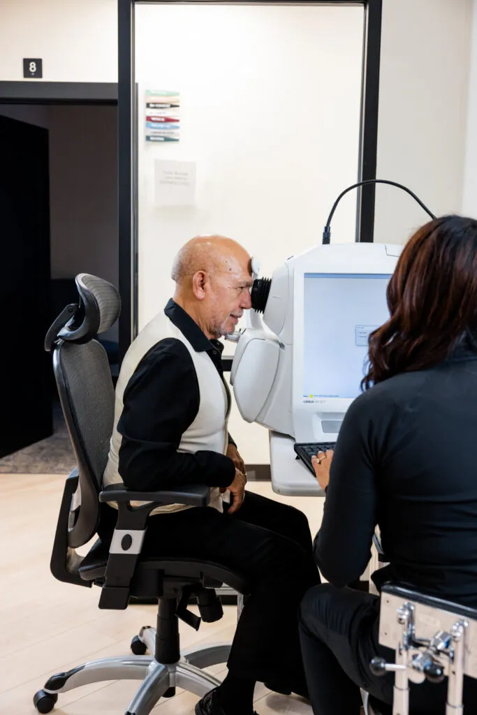
Optical Coherence Tomography Retinal Scan (OCT)
Light — not radiation — builds a cross-section of your retina layer by layer. Measuring the thickness of each layer lets your doctor detect retinal and optic-nerve disease long before it reaches your vision.
-
Uses Light, non-invasive
-
Shows Retinal layers
-
Detects Early disease
FIELD-OF-VIEW MAPPING
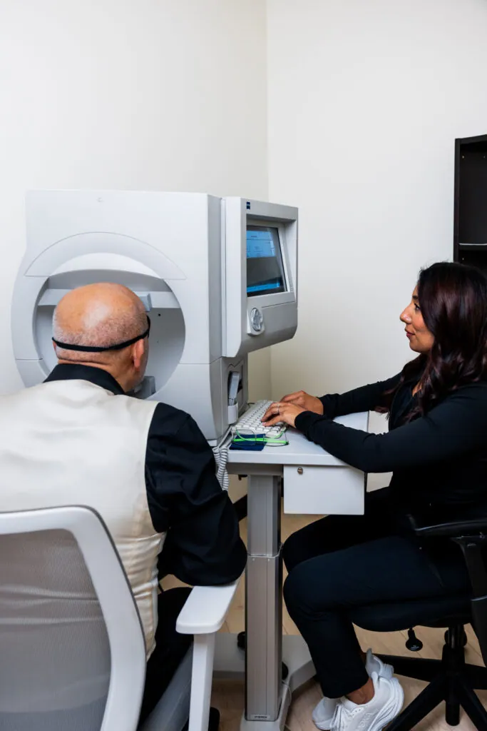
Visual Field
Points of light appear across your periphery and center to map exactly where you see — and where you don’t. It’s sensitive enough to reveal early signs of glaucoma, stroke, and neurological change.
-
Tests Periphery + center
-
Flags Glaucoma · stroke
-
Sensitivity Sub-clinical
CORNEAL MAPPING
Corneal Topographer
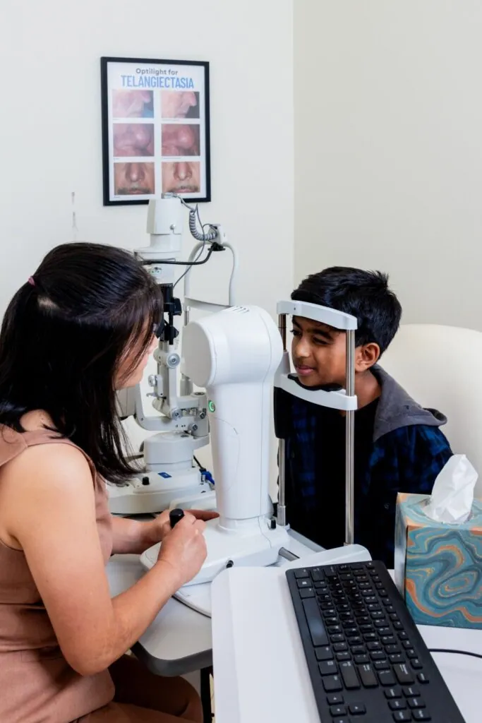
Thousands of measurement points map the shape of the front of your eye in a detailed color map. It is essential for fitting Ortho-K and specialty contact lenses, detecting conditions like keratoconus, and evaluating candidates for laser vision correction.
-
Maps Corneal shape
-
Used for Ortho-K & specialty lenses
-
Detects Keratoconus
ANTERIOR SEGMENT IMAGING
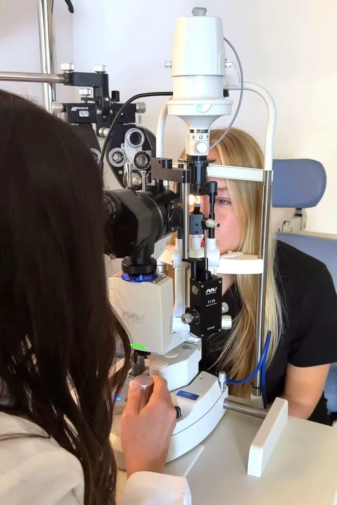
Firefly Slit-Lamp Video Camera
A high-definition camera on our slit lamp captures magnified photos and video of the front of your eye — eyelids, tear film, cornea and lens. Your doctor can show you exactly what they see, and compare images from one visit to the next to track any changes.
-
Views Front of the eye
-
Records HD photos & video
-
Helps Track changes
VILLAGE VISION CENTER
Come see what we see.
The place where care, comfort and community come together.